Current Books

Edited by: Justin D. Radolf and D. Scott Samuels Published: 2021
Book: 978-1-913652-61-6. Ebook: 978-1-913652-62-3
Indispensable for anyone involved in Borrelia and Lyme disease research and is highly recommended for microbiologists, immunologists, and physicians with an interest in spirochetes, vector-borne illness, or emerging infectious diseases read more ...
Full information at Lyme Disease and Relapsing Fever Spirochetes.
Edited by: Laurel J. Gershwin and Amelia R. Woolums Published: 2020
Book: 978-1-913652-59-3. Ebook: 978-1-913652-60-9
This concise book captures the essence of current and future shifts in vaccine development research that will likely transform our understanding of methods to stimulate specific and protective immune responses to infectious diseases read more ...
Full information at Veterinary Vaccines.

Edited by: Jürgen Marxsen Published: 2020
Book: 978-1-913652-57-9. Ebook: 978-1-913652-58-6
Climate change is continuing unabated and this new, expanded edition contains revised and updated chapters and the addition of four new chapters covering more of the topical fields in this important area of climate science. This is an essential book for every microbial ecologist read more ...
Full information at Climate Change and Microbial Ecology.

Edited by: Ekaterina E. Heldwein and Gregory A. Smith Published: 2020
Book: 978-1-913652-55-5. Ebook: 978-1-913652-56-2
This timely book is essential reading for everyone working on neuroinvasive alphaherpesviruses read more ...
Full information at Alphaherpesviruses.

Edited by: Jacob Moran-Gilad and Rachel E. Gibbs Published: 2020
Book: 978-1-913652-53-1. Ebook: 978-1-913652-54-8
This volume covers the epidemiology and ecology of Legionella, diagnosis and treatment of legionellosis and presents reviews of current and emerging concepts and new advances in Legionella research read more ...
Full information at Legionellosis Diagnosis and Control in the Genomic Era.
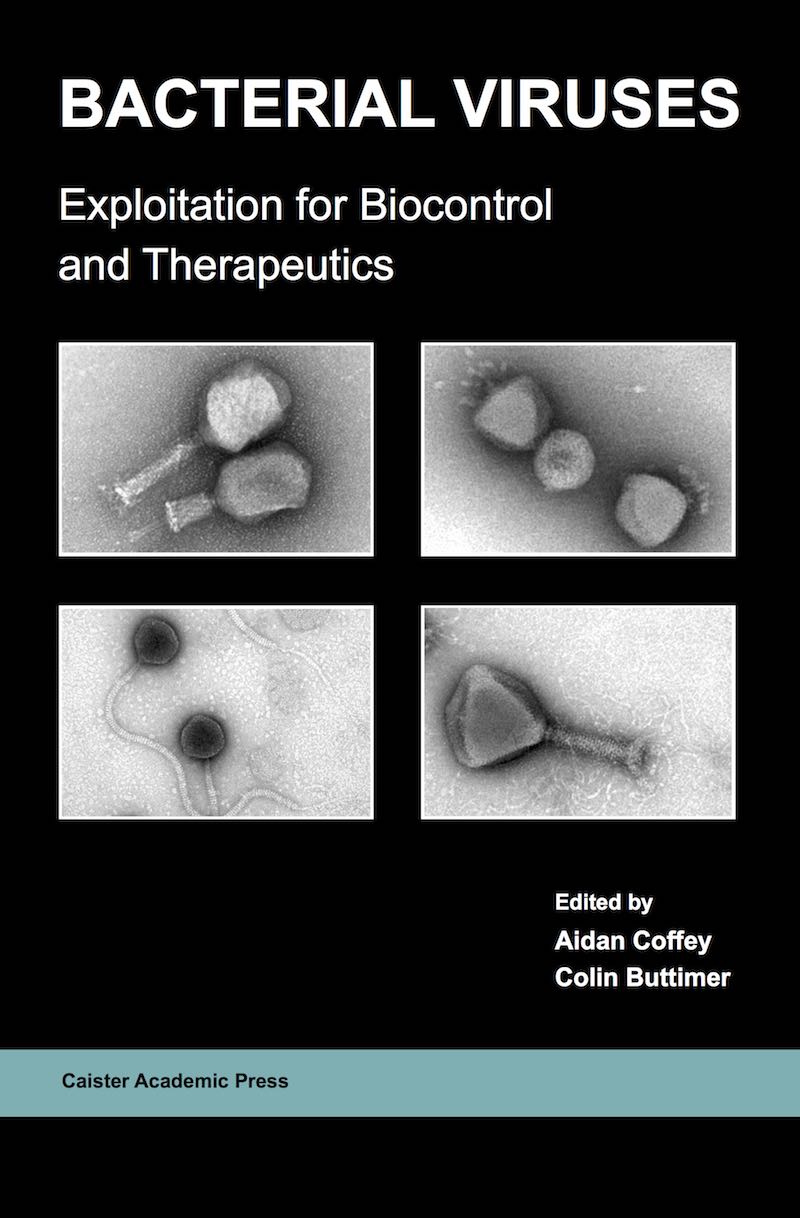
Edited by: Aidan Coffey and Colin Buttimer Published: 2020
Book: 978-1-913652-51-7. Ebook: 978-1-913652-52-4
Internationally-recognised scientists describe the exploitation of bacterial viruses in diverse areas including applications for biocontrol of undesirable bacteria in human and veterinary medicine, horticulture, aquaculture and food read more ...
Full information at Bacterial Viruses.
Edited by: Arindam Mitra Published: 2020
Book: 978-1-912530-32-8. Ebook: 978-1-912530-33-5
"for graduate students and researchers" (Ringgold); "accurate, up-to-date information ... a useful guide" (Doodys) read more ...
Full information at Microbial Biofilms.

Edited by: André Antunes Published: 2020
Book: 978-1-912530-30-4. Ebook: 978-1-912530-31-1
an up-to-date insight into current topics and research work ... a very good introduction to interested readers (BioSpektrum); "recent theoretical and experimental results" (Ringgold) read more ...
Full information at Astrobiology.
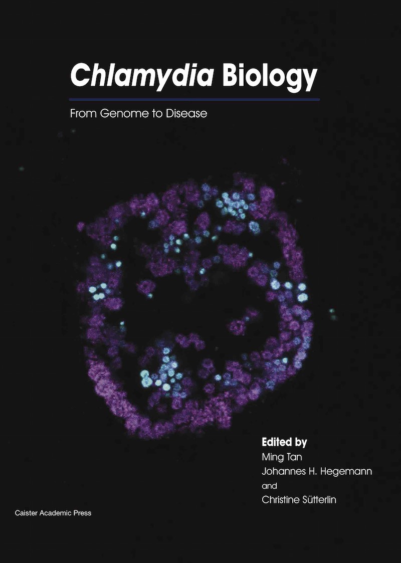
Edited by: Ming Tan, Johannes H. Hegemann and Christine Sütterlin Published: 2020
Book: 978-1-912530-28-1. Ebook: 978-1-912530-29-8
"The book as a whole is recommended to research students, doctoral students and scientists" (Biospektrum); "a current and comprehensive summary of Chlamydia research" (Doodys); "a broad reference on the bacterial pathogen Chlamydia and the human and animal disease it causes" (Ringgold) read more ...
Full information at Chlamydia Biology.
Edited by: Eugenia Corrales-Aguilar and Martin Schwemmle Published: 2020
Book: 978-1-912530-14-4. Ebook: 978-1-912530-15-1
"highly recommended" (Southeastern Naturalist) read more ...
Full information at Bats and Viruses.

Edited by: Van G. Wilson Published: 2019
Book: 978-1-912530-12-0. Ebook: 978-1-912530-13-7
"a comprehensive, in-depth resource ... intensive and technically detailed descriptions of the latest advances ... densely packed with information ... a valuable reference for any laboratory group working in this field" (Doodys) read more ...
Full information at SUMOylation and Ubiquitination.

Edited by: Siba K. Samal Published: 2019
Book: 978-1-912530-10-6. Ebook: 978-1-912530-11-3
"a nice introduction to avian virology" (Doodys); "this book is a must-have for anyone whose daily activities require detailed knowledge of the biology, pathogenesis, immune response, prevention, and control of avian viruses" (JAVMA) read more ...
Full information at Avian Virology.

Edited by: Özlem Ateş Duru Published: 2019
Book: 978-1-912530-26-7. Ebook: 978-1-912530-27-4
"of immense value for PhD students, postdoctorate students, microbiologists, and experienced scientists" (Doodys) read more ...
Full information at Microbial Exopolysaccharides.
Author: Mark A. Behlke, Kornelia Berghof-Jäger, Tom Brown, et al. Published: 2019
Book: 978-1-912530-24-3. Ebook: 978-1-912530-25-0
This indispensable manual is a compilation of review articles written by experts in the field of PCR technology read more ...
Full information at Polymerase Chain Reaction.

Edited by: Yuqing Li and Xuedong Zhou Published: 2019
Book: 978-1-912530-22-9. Ebook: 978-1-912530-23-6
Essential reading for everyone working with streptococci from the PhD student to the experienced scientist read more ...
Full information at Pathogenic Streptococci.
Edited by: Bryony C. Bonning Published: 2019
Book: 978-1-912530-08-3. Ebook: 978-1-912530-09-0
"essential reading for students and scholars of insect virology" (Biotechnol. Agron. Soc. Environ.); "I would recommend it to all researchers and students interested in insect viruses and advanced biotechnological applications" (Q. Rev. Biol.) read more ...
Full information at Insect Molecular Virology.

Edited by: Ludmila Chistoserdova Published: 2019
Book: 978-1-912530-04-5. Ebook: 978-1-912530-05-2
"highlights the diversity of methylotrophs, their functions, and their potential applications and will be of interest to many" (SIMB News) read more ...
Full information at Methylotrophs and Methylotroph Communities.

Edited by: Akikazu Sakudo and Takashi Onodera Published: 2019
Book: 978-1-910190-95-1. Ebook: 978-1-910190-96-8
"well written and comprehensive, and any research scientist who studies prion diseases should have this book in their reference library" (JAVMA) read more ...
Full information at Prions.

Edited by: Takashi Matsumoto and Yoshio Yamaoka, Published: 2019
Book: 978-1-910190-93-7. Ebook: 978-1-910190-94-4
"I would recommend this book" (Gut Microbes); "a very interesting book ... well written ... effective illustrations ... extensive reference list" (SIMB News) read more ...
Full information at Microbiota.

Edited by: Diana Marco Published: 2019
Book: 978-1-912530-02-1. Ebook: 978-1-912530-03-8
"easy to read ... applicable to teaching faculty as well as advanced undergraduate students, graduate students, and researchers" (SIMB News); "concise and well written" (Quarterly Rev. Biol.) read more ...
Full information at Microbial Ecology.