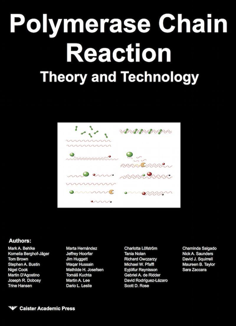
Real-Time PCR Technology: Fluorescence Detection
Since the fist scientific work published in 1996 the number of publications where real-time PCR is used has increased almost exponentially.
Rodríguez-Lázaro, D. and Hernández, M. (2013). Introduction to the Real-time PCR In Real-Time PCR in Food Science: Current Technology and Applications. D. Rodriguez-Lazaro, ed. (Norfolk, UK: Caister Academic Press). ISBN: 978-1-908230-15-7.
Real-time PCR Chemistries and Detection Formats
The fluorescence that is monitored during the entire real-time PCR process can be detected by a nonspecific detection strategy independent of the target sequence, e.g. through fluorescent dyes that have special fluorescent properties when bound to dsDNA, or by sequence-specific fluorescent oligonucleotide probes; i.e. a sequence-specific strategy (Rodríguez-Lázaro, D. and Hernández, M. 2013. Introduction to the Real-time PCR In Real-Time PCR in Food Science: Current Technology and Applications.)Nonspecific detection formats
Ethidium bromide was the first dye used for this purpose. Other intercalating dyes such as YO-PRO-1, BEBO or SYBR Green I have since been used. SYBR Green I is the most frequently used dsDNA-specific dye in real-time PCR. It is an asymmetric cyanine dye, structurally related to the dsDNA-specific dyes YOYO-1 and TOTO-1. Its binding affinity is more than 100 times higher than that of ethidium bromide. SYBR Green I largely binds sequence independently to the minor groove of ds-DNA. It can be excited with blue light with a wavelength of 480 nm, and its emission spectrum is comparable to that of fluorescein with a maximum at 520 nm and a quantum yield of 0.8. The fluorescence of the bound dye is above 1000-fold higher than that of the free dye and, therefore, is well suited for monitoring the product accumulation during PCR. When monitored in real time, this results in an increase in the fluorescence signal that can be observed during the polymerisation step, and that falls off when the DNA is denatured. Consequently, fluorescence measurements have to be performed at the end of the elongation step of every PCR cycle. This method obviates the need for target-specific fluorescent probes, and hence it can be used with any pair of primers for any target, making its use less expensive. However, its major disadvantage is that specificity is determined entirely by the primers and thus the risk of amplifying non-specific PCR products has to be considered during optimization. However, PCR product verification can be achieved at the end-point by plotting fluorescence as a function of temperature to generate a melting curve of the amplicon.The AmpliFluor system is an nonspecific detection system developed by Intergen co. AmpliFluor technology uses a universal energy-transfer hairpin primer (UniPrimer) which emits a fluorescent signal when unfolded during its incorporation into an amplification product. The UniPrimer contains a 18-nucleotides sequence (Z sequence: 5' -act gaa cct gac cgt aca-3') at its 3' end, that is also present at the 5' end of one of the target-specific primers so that it anneals to the PCR product and acts as universal PCR primer. In the first step, the forward primer is extended. This extended product serves as template for the reverse primer in the second step. In the end, the polymerase opens the hairpin structure and a double-stranded PCR product is formed in which reporter and quencher are separated. 3.2 Sequence-specific fluorescent oligonucleotide probes
There are different types of specific-sequence fluorescent probes, and they can be classified into two major groups: hydrolysis probes and hybridization probes, both types being homologous to the internal region amplified by the two primers. The fluorescence signal intensity can be related to the amount of PCR product (i) by a product-dependent decrease of the quench of a reporter fluorophore or (ii) by an increase of the fluorescence resonance energy transfer (FRET) from a donor to an acceptor fluorophore. FRET, also called Förster transfer, is the radiationless transfer of excitation energy by dipole-dipole interaction between fluorophores with overlapping emission and excitation spectra. The FRET and the quench efficiency are strongly dependent on the distance between the fluorophores. Therefore, the PCR-product-dependent change in the distance between the fluorophores is used to generate the sequence-specific signals. Several different formats can be used. In principle, all of them could function by a decrease of quench or an increase of FRET; in practice, most formats are based on a decrease of quench. The most commonly used fluorescent reporter dyes are FAM, TET (tetrachloro-6-car-boxyfluorescein), JOE (2,7-dimethoxy-4,5-dichloro-6-carboxy-fluorescein) or HEX (hexacholoro-6-carboxyfluorescein), and the most frequently used quenchers are TAMRA, DABCYL and Black Hole Quencher (BHQ).
Sequence-specific probes allow multiplexing and easy identification of point mutations. A common drawback of probe systems that use the decrease-of-quench mechanism is unwanted generation of a signal due to probe destruction (e.g. by unintentional hydrolysis of the probes by the Taq DNA polymerase) or by formation of secondary structures of the probes that lead to a decrease in quench.
Hydrolysis probes
The hydrolysis probes are cleaved when hybridised by the 5' - 3' exonuclease activity of particular DNA polymerases during the elongation phase of primers, yielding a real time measurable fluorescence emission directly proportional to the concentration of the target sequence. It usually utilises either Taq or Tth polymerase, but any enzyme with equivalent 5' - 3' exonuclease activity properties (e.g. Tfl) can be used. The best known hydrolysis probes are TaqMan® probes and TaqMan MGB (minor groove binder) probes, both developed by Applied Biosystems.A TaqMan probe is an oligonucleotide double-labelled with a reporter fluorophore at the 5' end (reporter dye) and with a quencher internally or at the 3' end (quencher dye). In addition, the probes must be blocked at their 3' - end to prevent the extension during the annealing step. The TaqMan assay uses three oligonucleotides. Two conventional primers allow amplification of the product, to which the TaqMan probe will anneal. The quencher dye absorbs the fluorescence of the reporter dye due to its proximity, which permits FRET. When the correct amplicon is amplified, the probe can hybridise to the target after the denaturation step. It remains hybridised while the polymerase extends the primers until it reaches the probe. Then, it displaces its 5' end to hold it in a forked structure. The enzyme continues to move from the now free end to the bifurcation of the duplex, where cleavage takes place. The quencher is hence released from the fluorophore, which now fluoresces after excitation. As the polymerase will cleave the probe only while it remains hybridised to its complementary strand, the temperature conditions of the polymerisation phase of the PCR must be adjusted to ensure probe binding. Most probes have a Tm of around 70 °C; therefore, the TaqMan system uses a combined annealing and polymerisation step at 60-63 °C. This ensures that the probe remains bound to its target during the primer extension step. It also ensures maximum 5' - 3' exonuclease activity of the Taq and Tth DNA polymerases.
The TaqMan MGB probes are similar to TaqMan probes. They contain a non-fluorescent quencher (NFQ) and an oligopeptide at the 3' end. This oligopeptide is a DNA minor groove binder (MGB), with very high affinity for the minor groove of A-T-rich double-stranded DNA. Addition of the MGB ligand significantly enhances duplex stability. The shorter the probe, the greater the MGB contribution to the overall duplex stability: with 12-18-bp oligonucleotides, it raises the Tm from 44-56°C up to 66-70°C. This allows designing suitable probes in sequences such as those rich in A-T, in which conventional TaqMan® probes require an excessive length.
Hybridization probes
In contrast to hydrolysis probes, hybridization probes are not hydrolyzed during PCR. The fluorescence is generated by a change in its secondary structure during the hybridization phase, which results in an increase of the distance separating the reporter and the quencher dyes. The most relevant hybridization probes are those containing hairpins (Molecular Beacons, Scorpion primers, etc), and FRET hybridization probes. Molecular beacons form a stem-and-loop structure through complementary sequences on the 5' and 3' ends of the probe. The loop portion is complementary to the target nucleic acid. A reporter and a quencher fluorophore are attached one at the end of each arm. The quencher is a non-fluorescent chromophore that dissipates the energy it receives from the fluorophore as heat. The fluorescence is quenched when the probe is in a hairpin-like structure (stem-and-loop structure) due to the proximity between quencher and fluorophore allowing FRET. In the presence of a complementary sequence, designed internal to the primer binding sites, the probe undergoes a conformational transition that forces the stem apart and results in the formation of a probe/target hybrid that is more stable than the former stem. This conformational change separates the fluorophore from the quencher and consequently FRET no longer occurs, thus increasing reporter fluorescence emission. Molecular Beacons have been reported to be significantly more specific than conventional oligonucleotide probes of equivalent length, due to the presence of a stem structure. The main drawback of Molecular Beacons is associated with its design as the fluorescence yield is very sensitive to the hybridisation conditions. FRET Probes or Hybridization probes use four oligonucleotides i.e. two primers and two sequence-specific probes. Each probe has a single label: either a donor fluorophore at the 3' -end or an acceptor fluorophore at the 5' -end. The emission spectrum of the donor fluorophore overlaps the excitation spectrum of the acceptor fluorophore. The FRET probes must be blocked at their 3' -end to prevent the extension during the annealing step. The two probes hybridize to the target sequences in a head-to-tail arrangement, thus bringing the two dyes close (typically 1-5 nucleotides distant), allowing FRET. During PCR, only the donor fluorophore is excited. In solution, only background fluorescence is emitted by the donor. During annealing, the two probes hybridise adjacently to their target sequence and thus the excitation energy is transferred by FRET from the donor dye in one of the probes to the acceptor dye in the other probe , allowing the acceptor dye to dissipate fluorescence at a different wavelength. The use of two independent probes results in high specificity and flexibility for probe design. Furthermore, as the probes are not hydrolysed, fluorescence is reversible and allows the generation of melting curves.Scorpion primers are structurally and functionally related to molecular beacons, but serve as primers in the PCR. They consist of a probe sequence linked to the 5' end of a primer via a non-amplifiable stopper moiety. The probe presents a fluorophore linked at the 5' -end and a quencher at the 3' -end, and is held in a hairpin loop structure by complementary sequences on its 5' - and 3' - ends. This configuration brings the fluorophore in close proximity with the quencher and avoids fluorescence similarly to Molecular Beacons. In addition, the probe sequence is complementary to an internal region of the sequence extended by the adjacent primer. In the first step, the primer is extended, yielding a single-stranded template for the reverse primer in the second step. Upon hybridization, the hairpin is opened, producing a physical separation of the fluorophore and quencher such that increases in signal are observed. In contrast to the sunrise primers, the reverse extension is blocked by a hexethylene glycol group. This ensures that the reporter of the scorpion primer remains quenched in nonspecific products like primer dimers.
The light-up technology utilizes a nucleic acid analogue instead of natural DNA as sequence recognizing element. Light-up probes are peptide nucleic acids (PNAs) that use thiazole orange, a derivative of cyanine, as reporter fluorophore. It forms sequence-specific complexes with DNA and RNA which are more stable than double-stranded natural nucleic acids. These features are attributed mainly to the charge-neutral nature of PNA, which eliminates the electrostatic repulsion between the hybridizing strands. The probe has low fluorescence when free in solution, however they show increased fluorescence intensity upon hybridisation with DNA.
Rodríguez-Lázaro, D. and Hernández, M. (2013). Introduction to the Real-time PCR In Real-Time PCR in Food Science: Current Technology and Applications. D. Rodriguez-Lazaro, ed. (Norfolk, UK: Caister Academic Press). ISBN: 978-1-908230-15-7.
Further reading
- Real-Time PCR: Advanced Technologies and Applications
- Real-Time PCR in Food Science: Current Technology and Applications
- Quantitative Real-time PCR in Applied Microbiology


